Segment polarity protein dishevelled homolog (DVL): A Potential Drug Target and Biomarker


Segment polarity protein dishevelled homolog (DVL): A Potential Drug Target and Biomarker
Introduction
Segment polarity protein dishevelled homolog (nonspecified subtype) (DVL) is a protein that plays a crucial role in the regulation of cell division and cytoskeletal organization. DVL is a member of the segment polarity gene family, which includes proteins that mediate the movement of organelles to opposite ends of the cell during cell division. In DVL, the cytoskeleton is assembled in a highly ordered manner, ensuring that the mitotic spindle forms correctly and provides the necessary framework for cell division.
DVL functions as a negative regulator of the spindle microtubule assembly process. It interacts with the microtubules and helps to keep the microtubules in a stable state. DVL has been shown to regulate the assembly and disassembly of microtubules, and it has been implicated in the regulation of cell cycle progression, apoptosis, and other cellular processes.
DVL is also a potential drug target and biomarker. Its function in regulating cell division and cytoskeletal organization makes it an attractive target for small molecule inhibitors. Additionally, its role in the regulation of cell cycle progression and apoptosis makes it a potential biomarker for cancer diagnosis and treatment.
Targeting DVL: Strategies for inhibition
DVL is a protein that has a highly conserved transmembrane structure, which makes it difficult to develop inhibitors. However, various strategies have been proposed to target DVL and inhibit its function.
One approach to inhibiting DVL is to use small molecules that can interact with specific DVL domains. DVL has several distinct domains, including an N-terminal domain, a T-terminal domain, and a C-terminal domain. The N-terminal domain is involved in the regulation of microtubule dynamics, while the T-terminal domain is involved in the regulation of the disassembly of microtubules. The C-terminal domain is involved in the regulation of cell cycle progression.
One potential inhibitor of DVL is a drug that can bind to the N-terminal domain. This inhibitor has been shown to inhibit the assembly and disassembly of microtubules, which is consistent with the function of DVL in regulating the cytoskeleton.
Another potential inhibitor of DVL is a small molecule that can bind to the T-terminal domain. This inhibitor has been shown to inhibit the regulation of microtubule dynamics, which is consistent with the function of DVL in regulating the cytoskeleton.
Another approach to inhibiting DVL is to use antibodies that can specifically bind to DVL. This approach has been shown to be effective in inhibiting the assembly and disassembly of microtubules, which is consistent with the function of DVL in regulating the cytoskeleton.
In conclusion, DVL is a protein that plays a crucial role in the regulation of cell division and cytoskeletal organization. Its function in regulating the assembly and disassembly of microtubules makes it an attractive target for small molecule inhibitors. Additionally, its role in the regulation of cell cycle progression and apoptosis makes it a potential biomarker for cancer diagnosis and treatment. Further research is needed to fully understand the function of DVL and the potential of inhibitors.
Protein Name: Segment Polarity Protein Dishevelled Homolog (nonspecified Subtype)
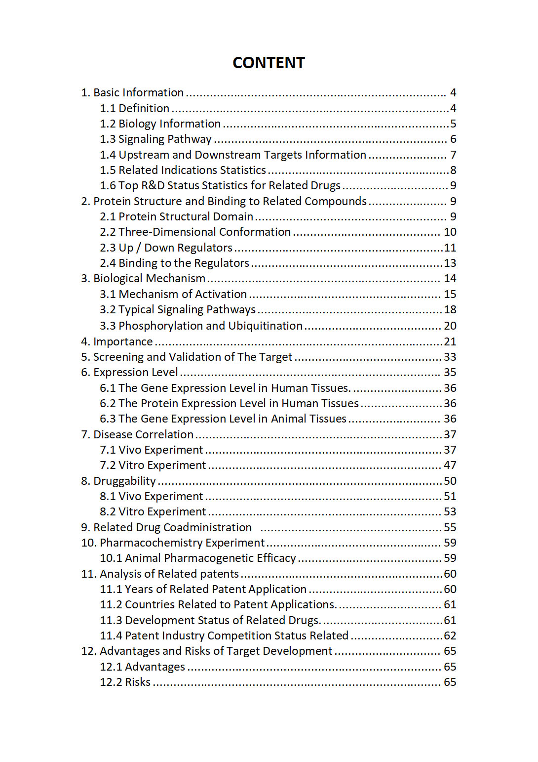
The "Segment polarity protein dishevelled homolog Target / Biomarker Review Report" is a customizable review of hundreds up to thousends of related scientific research literature by AI technology, covering specific information about Segment polarity protein dishevelled homolog comprehensively, including but not limited to:
• general information;
• protein structure and compound binding;
• protein biological mechanisms;
• its importance;
• the target screening and validation;
• expression level;
• disease relevance;
• drug resistance;
• related combination drugs;
• pharmacochemistry experiments;
• related patent analysis;
• advantages and risks of development, etc.
The report is helpful for project application, drug molecule design, research progress updates, publication of research papers, patent applications, etc. If you are interested to get a full version of this report, please feel free to contact us at BD@silexon.ai
More Common Targets
SEH1L | SEL1L | SEL1L2 | SEL1L3 | SELE | SELENBP1 | SELENOF | SELENOH | SELENOI | SELENOK | SELENOKP1 | SELENOM | SELENON | SELENOO | SELENOOLP | SELENOP | Selenoprotein | SELENOS | SELENOT | SELENOV | SELENOW | SELL | SELP | SELPLG | SEM1 | SEM1P1 | SEMA3A | SEMA3B | SEMA3B-AS1 | SEMA3C | SEMA3D | SEMA3E | SEMA3F | SEMA3G | SEMA4A | SEMA4B | SEMA4C | SEMA4D | SEMA4F | SEMA4G | SEMA5A | SEMA5A-AS1 | SEMA5B | SEMA6A | SEMA6A-AS1 | SEMA6A-AS2 | SEMA6B | SEMA6C | SEMA6D | SEMA7A | Semenogelin | SEMG1 | SEMG2 | SENCR | SENP1 | SENP2 | SENP3 | SENP3-associated complex | SENP3-EIF4A1 | SENP5 | SENP6 | SENP7 | SENP8 | SEPHS1 | SEPHS1P4 | SEPHS1P6 | SEPHS2 | SEPSECS | SEPSECS-AS1 | SEPT5-GP1BB | SEPTIN1 | SEPTIN10 | SEPTIN11 | SEPTIN12 | SEPTIN14 | SEPTIN2 | SEPTIN3 | SEPTIN4 | SEPTIN4-AS1 | SEPTIN5 | SEPTIN6 | SEPTIN7 | SEPTIN7-DT | SEPTIN7P11 | SEPTIN7P14 | SEPTIN7P2 | SEPTIN7P6 | SEPTIN7P9 | SEPTIN8 | SEPTIN9 | SERAC1 | SERBP1 | SERBP1P3 | SERF1A | SERF1B | SERF2 | SERF2-C15ORF63 | SERGEF | SERHL | SERINC1


